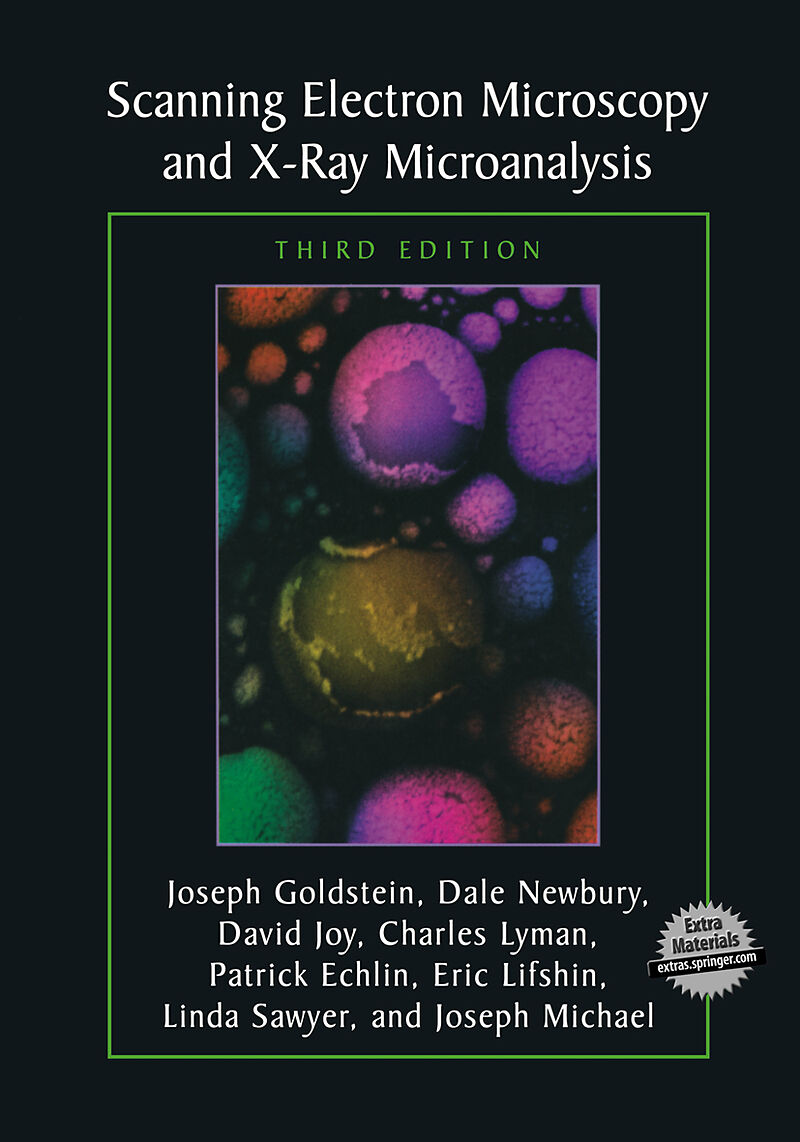
Scanning Electron Microscopy and X-Ray Microanalysis
Format:
E-Book (pdf)
EAN:
9781461502159
Untertitel:
Third Edition
Genre:
Technik
Autor:
Joseph Goldstein, Dale E. Newbury, David C. Joy, Charles E. Lyman, Patrick Echlin, Eric Lifshin, Linda Sawyer, J. R. Michael
Herausgeber:
Springer US
Anzahl Seiten:
689
Erscheinungsdatum:
06.12.2012
This text provides students as well as practitioners with a comprehensive introduction to the field of scanning electron microscopy (SEM) and X-ray microanalysis. The authors emphasize the practical aspects of the techniques described. Topics discussed include user-controlled functions of scanning electron microscopes and x-ray spectrometers and the use of x-rays for qualitative and quantitative analysis. Separate chapters cover SEM sample preparation methods for hard materials, polymers, and biological specimens. In addition techniques for the elimination of charging in non-conducting specimens are detailed.
Inhalt
1. Introduction.- 1.1. Imaging Capabilities.- 1.2. Structure Analysis.- 1.3. Elemental Analysis.- 1.4. Summary and Outline of This Book.- Appendix A. Overview of Scanning Electron Microscopy.- Appendix B. Overview of Electron Probe X-Ray Microanalysis.- References.- 2. The SEM and Its Modes of Operation.- 2.1. How the SEM Works.- 2.1.1. Functions of the SEM Subsystems.- 2.1.1.1. Electron Gun and Lenses Produce a Small Electron Beam.- 2.1.1.2. Deflection System Controls Magnification.- 2.1.1.3. Electron Detector Collects the Signal.- 2.1.1.4. Camera or Computer Records the Image.- 2.1.1.5. Operator Controls.- 2.1.2. SEM Imaging Modes.- 2.1.2.1. Resolution Mode.- 2.1.2.2. High-Current Mode.- 2.1.2.3. Depth-of-Focus Mode.- 2.1.2.4. Low-Voltage Mode.- 2.1.3. Why Learn about Electron Optics?.- 2.2. Electron Guns.- 2.2.1. Tungsten Hairpin Electron Guns.- 2.2.1.1. Filament.- 2.2.1.2. Grid Cap.- 2.2.1.3. Anode.- 2.2.1.4. Emission Current and Beam Current.- 2.2.1.5. Operator Control of the Electron Gun.- 2.2.2. Electron Gun Characteristics.- 2.2.2.1. Electron Emission Current.- 2.2.2.2. Brightness.- 2.2.2.3. Lifetime.- 2.2.2.4. Source Size, Energy Spread, Beam Stability.- 2.2.2.5. Improved Electron Gun Characteristics.- 2.2.3. Lanthanum Hexaboride (LaB6) Electron Guns.- 2.2.3.1. Introduction.- 2.2.3.2. Operation of the LaB6 Source.- 2.2.4. Field Emission Electron Guns.- 2.3. Electron Lenses.- 2.3.1. Making the Beam Smaller.- 2.3.1.1. Electron Focusing.- 2.3.1.2. Demagnification of the Beam.- 2.3.2. Lenses in SEMs.- 2.3.2.1. Condenser Lenses.- 2.3.2.2. Objective Lenses.- 2.3.2.3. Real and Virtual Objective Apertures.- 2.3.3. Operator Control of SEM Lenses.- 2.3.3.1. Effect of Aperture Size.- 2.3.3.2. Effect of Working Distance.- 2.3.3.3. Effect of Condenser Lens Strength.- 2.3.4. Gaussian Probe Diameter.- 2.3.5. Lens Aberrations.- 2.3.5.1. Spherical Aberration.- 2.3.5.2. Aperture Diffraction.- 2.3.5.3. Chromatic Aberration.- 2.3.5.4. Astigmatism.- 2.3.5.5. Aberrations in the Objective Lens.- 2.4. Electron Probe Diameter versus Electron Probe Current.- 2.4.1. Calculation of dmin and imax.- 2.4.1.1. Minimum Probe Size.- 2.4.1.2. Minimum Probe Size at 10-30 kV.- 2.4.1.3. Maximum Probe Current at 10-30 kV.- 2.4.1.4. Low-Voltage Operation.- 2.4.1.5. Graphical Summary.- 2.4.2. Performance in the SEM Modes.- 2.4.2.1. Resolution Mode.- 2.4.2.2. High-Current Mode.- 2.4.2.3. Depth-of-Focus Mode.- 2.4.2.4. Low-Voltage SEM.- 2.4.2.5. Environmental Barriers to High-Resolution Imaging.- References.- 3. Electron Beam-Specimen Interactions.- 3.1. The Story So Far.- 3.2. The Beam Enters the Specimen.- 3.3. The Interaction Volume.- 3.3.1. Visualizing the Interaction Volume.- 3.3.2. Simulating the Interaction Volume.- 3.3.3. Influence of Beam and Specimen Parameters on the Interaction Volume.- 3.3.3.1. Influence of Beam Energy on the Interaction Volume.- 3.3.3.2. Influence of Atomic Number on the Interaction Volume.- 3.3.3.3. Influence of Specimen Surface Tilt on the Interaction Volume.- 3.3.4. Electron Range: A Simple Measure of the Interaction Volume.- 3.3.4.1. Introduction.- 3.3.4.2. The Electron Range at Low Beam Energy.- 3.4. Imaging Signals from the Interaction Volume.- 3.4.1. Backscattered Electrons.- 3.4.1.1. Atomic Number Dependence of BSE.- 3.4.1.2. Beam Energy Dependence of BSE.- 3.4.1.3. Tilt Dependence of BSE.- 3.4.1.4. Angular Distribution of BSE.- 3.4.1.5. Energy Distribution of BSE.- 3.4.1.6. Lateral Spatial Distribution of BSE.- 3.4.1.7. Sampling Depth of BSE.- 3.4.2. Secondary Electrons.- 3.4.2.1. Definition and Origin of SE.- 3.4.2.2. SE Yield with Primary Beam Energy.- 3.4.2.3. SE Energy Distribution.- 3.4.2.4. Range and Escape Depth of SE.- 3.4.2.5. Relative Contributions of SE1 and SE2.- 3.4.2.6. Specimen Composition Dependence of SE.- 3.4.2.7. Specimen Tilt Dependence of SE.- 3.4.2.8. Angular Distribution of SE.- References.- 4. Image Formation and Interpretation.- 4.1. The Story So Far.- 4.2. The Basic SEM Imaging Process.- 4.2.1. Scanning Action.- 4.2.2. Image Construction (Mapping).- 4.2.2.1. Line Scans.- 4.2.2.2. Image (Area) Scanning.- 4.2.2.3. Digital Imaging: Collection and Display.- 4.2.3. Magnification.- 4.2.4. Picture Element (Pixel) Size.- 4.2.5. Low-Magnification Operation.- 4.2.6. Depth of Field (Focus).- 4.2.7. Image Distortion.- 4.2.7.1. Projection Distortion: Gnomonic Projection.- 4.2.7.2. Projection Distortion: Image Foreshortening.- 4.2.7.3. Scan Distortion: Pathological Defects.- 4.2.7.4. Moiré Effects.- 4.3. Detectors.- 4.3.1. Introduction.- 4.3.2. Electron Detectors.- 4.3.2.1. Everhart-Thornley Detector.- 4.3.2.2. "Through-the-Lens" (TTL) Detector.- 4.3.2.3. Dedicated Backscattered Electron Detectors.- 4.4. The Roles of the Specimen and Detector in Contrast Formation.- 4.4.1. Contrast.- 4.4.2. Compositional (Atomic Number) Contrast.- 4.4.2.1. Introduction.- 4.4.2.2. Compositional Contrast with Backscattered Electrons.- 4.4.3. Topographic Contrast.- 4.4.3.1. Origins of Topographic Contrast.- 4.4.3.2. Topographic Contrast with the Everhart-Thornley Detector.- 4.4.3.3. Light-Optical Analogy.- 4.4.3.4. Interpreting Topographic Contrast with Other Detectors.- 4.5. Image Quality.- 4.6. Image Processing for the Display of Contrast Information.- 4.6.1. The Signal Chain.- 4.6.2. The Visibility Problem.- 4.6.3. Analog and Digital Image Processing.- 4.6.4. Basic Digital Image Processing.- 4.6.4.1. Digital Image Enhancement.- 4.6.4.2. Digital Image Measurements.- References.- 5. Special Topics in Scanning Electron Microscopy.- 5.1. High-Resolution Imaging.- 5.1.1. The Resolution Problem.- 5.1.2. Achieving High Resolution at High Beam Energy.- 5.1.3. High-Resolution Imaging at Low Voltage.- 5.2. STEM-in-SEM: High Resolution for the Special Case of Thin Specimens.- 5.3. Surface Imaging at Low Voltage.- 5.4. Making Dimensional Measurements in the SEM.- 5.5. Recovering the Third Dimension: Stereomicroscopy.- 5.5.1. Qualitative Stereo Imaging and Presentation.- 5.5.2. Quantitative Stereo Microscopy.- 5.6. Variable-Pressure and Environmental SEM.- 5.6.1. Current Instruments.- 5.6.2. Gas in the Specimen Chamber.- 5.6.2.1. Units of Gas Pressure.- 5.6.2.2. The Vacuum System.- 5.6.3. Electron Interactions with Gases.- 5.6.4. The Effect of the Gas on Charging.- 5.6.5. Imaging in the ESEM and the VPSEM.- 5.6.6. X-Ray Microanalysis in the Presence of a Gas.- 5.7. Special Contrast Mechanisms.- 5.7.1. Electric Fields.- 5.7.2. Magnetic Fields.- 5.7.2.1. Type 1 Magnetic Contrast.- 5.7.2.2. Type 2 Magnetic Contrast.- 5.7.3. Crystallographic Contrast.- 5.8. Electron Backscatter Patterns.- 5.8.1. Origin of EBSD Patterns.- 5.8.2. Hardware for EBSD.- 5.8.3. Resolution of EBSD.- 5.8.3.1. Lateral Spatial Resolution.- 5.8.3.2. Depth Resolution.- 5.8.4. Applications.- 5.8.4.1. Orientation Mapping.- 5.8.4.2. Phase Identification.- References.- 6. Generation of X-Rays in the SEM Specimen.- 6.1. Continuum X-Ray Production (Bremsstrahlung).- 6.2. Characteristic X-Ray Production.- 6.2.1. Origin.- 6.2.2. Fluorescence Yield.- 6.2.3. Electron Shells.- 6.2.4. Energy-Level Diagram.- 6.2.5. Electron Transitions.- 6.2.6. Critical Ionization Energy.- 6.2.7. Moseley's Law.- 6.2.8. Families of Characteristic Lines.- 6.2.9. Natural Width of Characteristic X-Ray Lines.- 6.2.10. Weights of Lines.- 6.2.11. Cross Section for Inner Shell Ion…

Leider konnten wir für diesen Artikel keine Preise ermitteln ...
billigbuch.ch sucht jetzt für Sie die besten Angebote ...
Die aktuellen Verkaufspreise von 3 Onlineshops werden in Realtime abgefragt.
Sie können das gewünschte Produkt anschliessend direkt beim Anbieter Ihrer Wahl bestellen.
Loading...
Die aktuellen Verkaufspreise von 3 Onlineshops werden in Realtime abgefragt.
Sie können das gewünschte Produkt anschliessend direkt beim Anbieter Ihrer Wahl bestellen.
| # | Onlineshop | Preis CHF | Versand CHF | Total CHF | ||
|---|---|---|---|---|---|---|
| 1 | Seller | 0.00 | 0.00 | 0.00 |
